
Navicular Stress Fractures
Updated: Jul 30
In this article
What is a navicular fracture?
Why it gets missed
Who gets it and why
Symptoms to watch for
Diagnosis and imaging
Treatment at SportsFit
Recovery timelines
Book an appointment
Navicular stress fractures have a reputation for being missed — and it's a well-earned one. The pain is often vague, the location is easy to confuse with other foot conditions, and because it tends to come on gradually rather than with a single traumatic event, athletes frequently try to train through it for weeks or even months before getting a proper diagnosis.
The problem is that delayed diagnosis is the biggest driver of poor outcomes with navicular fractures. Get on top of it early and you're looking at a structured but manageable recovery. Ignore it for a season and you're risking a complete fracture, avascular necrosis of the bone, and potentially surgical intervention.
At SportsFit Health & Rehab in Five Dock and Gladesville, we treat navicular stress fractures in runners, cricketers, and field sport athletes across the Inner West and Northern Sydney. This guide covers everything you need to know — including why it gets missed and what proper management actually looks like.
What is a navicular stress fracture?
The navicular is a small, boat-shaped bone sitting at the top of the midfoot — part of the midtarsal joint complex and a critical load-bearing structure in the foot's medial arch. During walking and running, it acts as the keystone of the arch, absorbing and transmitting significant compressive and tensile forces with every step.
A stress fracture occurs when repetitive loading exceeds the bone's capacity to remodel and repair itself. Unlike an acute fracture from a single event, a stress fracture develops gradually — microdamage accumulates faster than the bone can heal, eventually producing a crack within the bone's structure.
The navicular is particularly vulnerable to this process for two reasons:
Poor blood supply to the central third
The navicular has a watershed zone — a region of poor vascularity in its central portion where blood supply from both sides of the bone converges but is least concentrated. This is exactly where most stress fractures occur. Poor blood supply means slower remodelling, slower healing, and higher risk of non-union if the fracture isn't managed properly.
High compressive load concentration
The navicular sits at the apex of the medial longitudinal arch. In running and jumping, ground reaction forces are funnelled through this point repeatedly — thousands of times per training session. In athletes with higher arches, a rigid midfoot, or poor ankle dorsiflexion, the concentration of force on the navicular increases further.
Stress fracture vs. acute fracture: This article focuses on navicular stress fractures — the most common navicular injury in athletes. Acute navicular fractures from trauma (a fall, a direct impact, or a severe midfoot sprain) are a different entity and require urgent imaging and orthopaedic assessment. If you've had a significant foot injury with immediate severe pain and swelling, that's a different conversation.
Why does it get missed?
Navicular stress fractures are notoriously under-diagnosed, and there are several reasons for this:
The pain is often non-specific. Athletes typically describe a vague ache across the top of the midfoot or into the medial arch — easy to confuse with plantar fasciitis, posterior tibial tendinopathy, or a general midfoot overuse complaint
It often hurts more at the start of activity, then eases. This pattern leads athletes to believe it's "just tightness" or a minor strain that will resolve with more warm-up. It won't — it'll get worse
X-rays frequently miss it. Plain radiographs are insensitive for navicular stress fractures — studies suggest X-ray misses approximately 30–40% of confirmed fractures. Normal X-ray does not rule it out
There's no clear traumatic event. With no single moment to point to, athletes and clinicians alike can underestimate the severity of what's happening
The gold standard for diagnosis is MRI or CT scan. MRI detects bony oedema (the early stress reaction stage before a frank fracture is present) and shows the extent of marrow involvement. CT scan better characterises the fracture line and is useful for surgical planning in complete fractures. Both are superior to plain X-ray by a significant margin.
Who gets navicular stress fractures, and why?
Runners
High weekly mileage, recent training load increases, or a return from injury with too-rapid volume build-up are the classic patterns. Runners with a high arch (cavus foot) are at particular risk due to the reduced shock absorption through the midfoot and concentrated loading at the navicular. Trail runners changing to road and track athletes switching to spikes are also common presentations.
Cricketers
Fast bowlers are disproportionately affected — the front foot impact during the delivery stride generates very high ground reaction forces through the midfoot, repeated hundreds of times through a bowling spell and over a full season. Cricket is actually one of the sports with the highest navicular stress fracture incidence relative to its participation numbers. We see this regularly in our Premier Grade cricket patient base.
Field sport athletes
AFL players, football players, and basketball athletes involved in repetitive cutting, jumping, and sprinting accumulate similar midfoot loading. The combination of hard playing surfaces and high session volumes during pre-season or condensed fixture schedules is a common background.
Contributing biomechanical factors
Limited ankle dorsiflexion range, a rigid or high-arched foot, leg length discrepancy, and poor single leg stability all alter how force is distributed through the midfoot. These don't cause the fracture on their own, but they increase the risk significantly when combined with high training loads.
Symptoms and what to look for
Typical presentation:
Gradual onset pain across the top of the midfoot — often described as a dull ache that worsens with activity
Pain that is worse at the start of activity, may ease mid-session, then returns and worsens after
Localised tenderness directly over the navicular bone (the bony prominence on the inner top of the midfoot)
Increasing pain over weeks, often misinterpreted as a soft tissue strain that "isn't getting better"
In later stages, pain at rest and significant pain with any weight-bearing activity
The "N spot" test:
Direct, firm palpation over the navicular — the "N spot" — that reproduces sharp pain is highly specific for navicular stress fracture. This is one of the most reliable clinical signs and should always prompt imaging. If you press firmly on the top of your midfoot just in front of the ankle joint and it produces sharp, localised pain, get it scanned — don't keep training on it.
Signs warranting urgent assessment:
Positive N spot test with a history of increasing midfoot pain in an active athlete — imaging required before continuing sport
Any athlete who has had a navicular stress fracture previously — risk of recurrence at the same site is significant
Significant swelling and bruising over the midfoot after trauma — rule out acute fracture
Inability to hop on one leg without significant pain — this is a useful field test that suggests a high-grade stress injury
Diagnosis and imaging
Our assessment at Five Dock or Gladesville will confirm the clinical picture and determine the appropriate imaging pathway:
Clinical examination — N spot palpation, single leg hop test, foot posture assessment, ankle dorsiflexion range, and load assessment
Training load history — recent mileage, session frequency, surface changes, footwear, return-from-injury status
Referral for MRI — our first-line imaging recommendation for suspected navicular stress fracture. Detects stress reactions before a frank fracture is visible on CT and gives the clearest picture of bony oedema extent
CT scan — if MRI confirms a fracture and we need to understand the fracture line and degree of cortical disruption for management planning. Essential for any complete fracture or when surgical input is being sought
We will not attempt to manage a suspected navicular stress fracture conservatively without imaging confirmation. The stakes are too high — a mismanaged navicular fracture can progress to non-union, collapse, or avascular necrosis, any of which dramatically extends the recovery timeline and may require surgery.
Treatment: what evidence-based navicular management looks like
Management depends on the grade of the stress injury on imaging. The Khan classification (Grade 1–3 on CT) is widely used to guide decisions, with Grade 1 representing a dorsal cortex fracture only, Grade 2 involving the fracture line propagating into the navicular body, and Grade 3 representing a complete fracture.
Non-weight-bearing and immobilisation (weeks 0–6)
For confirmed navicular stress fractures, the evidence consistently supports a period of non-weight-bearing in a below-knee cast or boot — typically 6 weeks for Grade 1–2 injuries. This is non-negotiable and is the single biggest factor in determining whether the fracture heals cleanly.
Athletes often push back on this — understandably, because six weeks non-weight-bearing is a significant commitment. But the alternative — attempting to manage with reduced loading while still bearing weight — has a substantially higher rate of delayed union, non-union, and re-fracture. The evidence is clear on this.
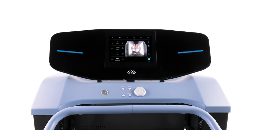
During the non-weight-bearing phase, maintaining cardiovascular fitness and upper body strength is important. At SportsFit Five Dock, we use the BTL R Force Anti-Gravity Treadmill as part of our management for appropriate navicular fracture patients — once cleared by the treating clinician and depending on the fracture grade, the anti-gravity treadmill allows athletes to walk and even jog at a fraction of their bodyweight (sometimes as low as 20%), maintaining cardiovascular fitness, movement quality, and lower limb neuromuscular activation while virtually eliminating the ground reaction force through the healing navicular. This makes a genuine difference to both physical conditioning and athlete wellbeing during what is otherwise a frustrating period of enforced rest.
Progressive weight-bearing return (weeks 6–10)
Once imaging shows evidence of healing and the athlete is clinically asymptomatic on palpation, a graduated return to weight-bearing begins:
Transition from non-weight-bearing to partial weight-bearing in a boot, progressing to full weight-bearing in the boot over 1–2 weeks
Weaning from the boot to supportive footwear based on symptom response
Foot and ankle strengthening — intrinsic foot muscles, peroneal strengthening, and calf loading beginning once full weight-bearing is comfortable
Hip and proximal strengthening — maintaining kinetic chain strength during the immobilisation period and building back on it
Gait retraining — returning to normal walking mechanics before any return to running is considered
For patients with significant deconditioning from the non-weight-bearing phase, we continue to use the anti-gravity treadmill to bridge the gap — allowing progressive loading of the foot and ankle in a controlled, low-impact environment before the athlete is ready for full ground reaction forces.
Shockwave therapy as an adjunct
For navicular stress fractures with delayed union or in the later stages of bone healing, extracorporeal shockwave therapy (ESWT) using our BTL-6000 device has emerging evidence as a useful adjunct. Shockwave stimulates osteoblast activity and promotes angiogenesis — essentially accelerating the biological processes involved in bone remodelling and vascularisation. While not a first-line treatment for acute navicular fractures, it can be a valuable tool in cases where healing is progressing slower than expected.
Surgical management
Grade 3 complete fractures, fractures that fail to unite with conservative management, or presentations with avascular necrosis of the navicular fragment may require surgical intervention — typically internal fixation with a screw. We work with orthopaedic colleagues in the Sydney area and facilitate referral when the clinical picture warrants it. Post-surgical physiotherapy follows a similar staged progression to conservative management but with timelines guided by the surgeon's protocol.
Return to running and sport (weeks 10–20+)
Return to running is milestone-driven, not time-driven. Before any running begins, we need:
Pain-free single leg hop — 10 consecutive hops without pain on the injured side
Full ankle dorsiflexion range
Symmetrical calf and foot intrinsic strength
Confirmed imaging healing (CT or MRI showing bridging callus or fracture resolution)
Return to running follows a walk-jog progression over 4–6 weeks, with careful monitoring of any symptoms at the navicular. Running volume builds at no more than 10% per week. Return to sport-specific training — bowling for cricketers, full field sport training for AFL and football players — comes after straight-line running is well established and symptom-free.
For our cricket patients, the bowling return is managed particularly carefully — the front foot impact during delivery is one of the highest-load events the navicular will face, and it's introduced last in the return-to-cricket protocol. We reference our cricket-specific rehabilitation approach to ensure the return program accounts for the unique demands of fast bowling.
Recovery timelines
Grade 1 — Non-weight-bearing period: 6 weeks; Return to running: 10–12 weeks; Return to full sport: 14–18 weeks
Grade 2 — Non-weight-bearing period: 6–8 weeks; Return to running: 12–16 weeks; Return to full sport: 16–24 weeks
Grade 3 / surgical — Non-weight-bearing period: 6–12 weeks; Return to running: 16–20+ weeks; Return to full sport: 6–12 months
These timelines assume imaging-confirmed healing progression and adherence to the non-weight-bearing protocol. Athletes who attempt to shortcut the immobilisation phase — returning to partial loading before the fracture has consolidated — consistently have worse outcomes, longer recovery times, and higher re-fracture rates.
Delayed union (fracture not healing as expected at the 6-week imaging check) adds 4–8 weeks to the timeline and may trigger a surgical discussion depending on the degree of non-union.
Reducing the risk of navicular stress fracture
Build training load gradually. The 10% rule for weekly mileage increase is a reasonable starting point — but individual tolerance varies. Athletes returning from injury or a break are at highest risk during the first 4–6 weeks of resumed training.
Address ankle dorsiflexion restriction. Limited ankle mobility increases midfoot loading. Regular calf and ankle mobility work, and assessment of whether footwear or orthotics are contributing, can make a meaningful difference.
Don't ignore persistent midfoot pain. A midfoot ache that isn't resolving after 2–3 weeks of reduced training should be assessed. Early-stage stress reactions (Grade 1 on MRI, no visible fracture line) can sometimes be managed with activity modification and monitoring rather than full immobilisation — but you won't know that without imaging.
Optimise foot mechanics. Custom orthotics may reduce navicular loading in athletes with high arches or significant overpronation. This is worth discussing with your physio if you've had a navicular fracture or have repeated midfoot complaints.
Previous fracture = higher risk. The navicular is prone to fracture at the same site. Athletes who've had one should be monitored more closely during load spikes and are strong candidates for preventive assessment at the start of each season.
Midfoot pain that won't go away?
If you've had persistent top-of-foot pain during or after running, cricket, or field sport — particularly if it's been going on for more than 2–3 weeks — don't keep training through it. Our team at SportsFit in Five Dock and Gladesville can assess it properly and get the right imaging arranged if needed.
Five Dock (Flagship) — Great North Road, Five Dock NSW
Gladesville — Gladesville NSW
**Same-day appointments available. Book online or call us directly.**


Comments