The Anatomy and Function of the Hip Joint: A Comprehensive Overview
- Sachin Bhat

- Nov 19, 2024
- 3 min read
Anatomy of the Hip Joint
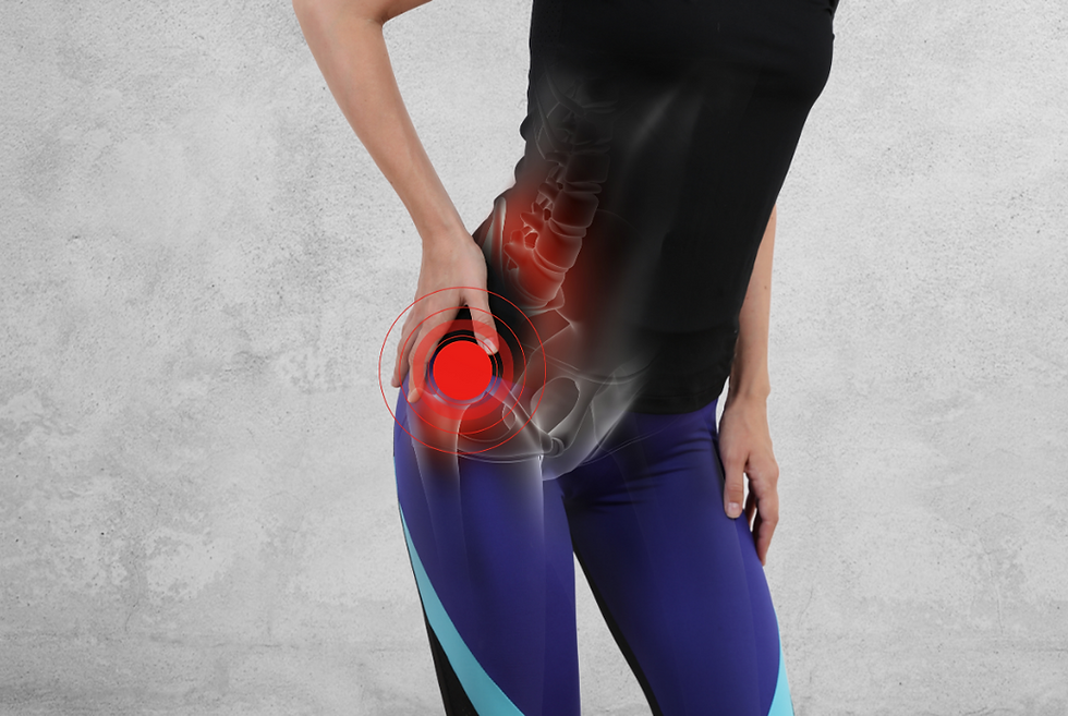
The hip joint is a critical part of our lower limb musculoskeletal system, playing a central role in supporting our body weight, enabling movement, and maintaining stability. For physiotherapists, understanding the anatomy of the hip is crucial for diagnosing and treating issues related to movement, strength, and joint pain. This post will explore the anatomy of the hip joint, including its bones, muscles, ligaments, and other key structures, to highlight how each component contributes to optimal hip function.

The Structure of the Hip Joint
The hip joint is a ball-and-socket synovial joint, one of the most stable and flexible joints in the body. It connects the femur (thigh bone) to the pelvis, and is designed to allow for a large range of motion.
- The Ball: The head of the femur (the rounded top of the thigh bone) forms the “ball” of the joint.
- The Socket: The acetabulum, a concave surface on the pelvis, forms the “socket” that accommodates the femoral head. The acetabulum is deepened by a rim of fibrocartilage known as the labrum, which provides additional stability and shock absorption, covering the femoral head by approximately 40% at any time.

Ligaments Supporting the Hip Joint
Ligaments around the hip joint play a critical role in stabilizing it, especially when we stand or move. Key ligaments include:
- Iliofemoral Ligament: One of the strongest ligaments in the body, it resists excessive extension and external rotation.
- Pubofemoral Ligament: This ligament prevents excessive abduction and extension, helping protect the hip against extreme movement.
- Ischiofemoral Ligament: Positioned at the back of the hip, this ligament wraps around the front of the joint to restrict internal rotation and extension.
Together, these ligaments provide a strong capsule around the joint, ensuring stability even under heavy loads.
Other Structures
- Labrum: The acetabular labrum is a ring of cartilage that deepens the hip socket, enhancing stability and reducing friction.
- Bursae: Small fluid-filled sacs called bursae are present around the hip joint to cushion and reduce friction between bones, tendons, and muscles.
- Capsule: Surrounds the joint to encase the synovial fluid, and enable fluid movement

Movement
The bony and soft tissue anatomy of the hip joint allows for a large range of motion to occur at the joint. The individual movements the hip joint is able to perform are flexion, extension, abduction, adduction, internal rotation, and external rotation, of which normal ranges are outlined below:
● Flexion – 120 degrees
● Extension – 10 degrees
● Abduction – 45 degrees
● Adduction – 25 degrees
● Internal Rotation – 15 degrees
● External rotation – 35 degrees
Clinical Relevance for Physiotherapy
The musculoskeletal anatomy of the hip joint, how it functions, and the type of structures involved informs the injuries that are more likely to occur here. For example, due to the strength of the ligaments around the hip, it is rare to have a hip ligamentous injury, and additionally due to the strong negative pressure within the hip joint, it is rare to have a hip joint dislocation. More common injuries that can occur at the hip joint include:
- Femoroacetabular impingement (FAI): bony changes to either the femur or acetabulum that results in bony restriction of the joint
- Labral tear: tear or affect to the acetabular labrum on the circumference of the hip joint, that may result in clicking, catching, or locking sensation of the hip joint
- Osteoarthritis: inflammation and reduction in space within the hip joint that may be a result of prior injury, or age
The hip joint’s complexity is what enables it to provide strength, stability, and mobility simultaneously. For physiotherapists, understanding each anatomical structure and its function is essential to diagnosing and understanding hip issues and developing treatment plans that effectively restore movement, reduce pain, and prevent future injuries.


Comments